
In the American healthcare system, how do diagnostic imaging systems help with early disease detection?
Diagnostic imaging devices are critical in the early detection of disease in the American healthcare system. These technologies enable clinicians to visualize the inside of the body non-invasively and spot irregularities that may signal the presence of disease. Diagnostic imaging can improve health outcomes and lower healthcare costs by detecting disorders at an earlier stage. Let us look at how various diagnostic imaging system methods are employed in the United States and how they contribute to early disease diagnosis.
X-ray Examination
X-ray imaging is one of the most extensively utilized and cost-effective diagnostic imaging modalities. Ionizing radiation in the form of X-rays can travel through biological tissues and be caught on photographic film or digital detectors. This enables doctors to view within bones and inside organs. Chest X-rays to check for lung abnormalities, mammograms to check for breast cancer, and dental X-rays are all common X-ray tests. X-rays for lung problems, mammograms for breast cancer screening, and dental x-rays. X-rays are extremely valuable for early detection of bone fractures, joint disorders, lung infections such as pneumonia, and some malignancies. Mammography screening programs, for example, have resulted in a drop in breast cancer mortality because tumors are now detected while they are smaller and more curable.
Imaging Using Ultrasound
Sonography, or ultrasound imaging, employs high-frequency sound waves to visualize soft tissues and organs inside the body. It is a non-ionizing radiation imaging technology that is extremely safe. During pregnancy, ultrasound is routinely used to monitor fetal development and detect abnormalities. It is also used to picture the abdomen to check for issues with organs such as the liver, gallbladder, pancreas, and kidneys. Ultrasound can detect gallstones, kidney stones, thyroid nodules, and soft tissue and organ abnormalities in their early stages. Ultrasound screening of the thyroid gland, for example, has resulted in the detection of more early-stage thyroid malignancies.
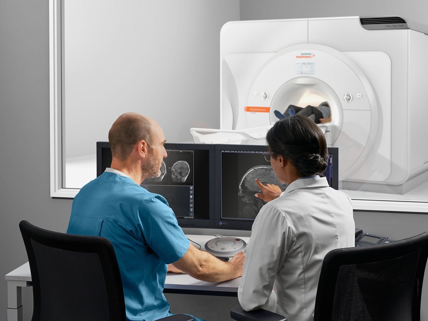
Scanning with Computed Tomography (CT)
CT scanning, also known as computed tomography or CAT scanning, creates cross-sectional images of the body using rotating X-ray beams and sophisticated computers. These precise images enable doctors to view bones and soft tissues. CT scans can detect and characterize abnormalities within organs such as the lungs, liver, pancreas, and colon. They’re also quite good at imaging the brain and detecting early signs of stroke. CT scans are useful for cancer screening and detecting malignancies at an early stage.
Magnetic Resonance Imaging (MRI)
MRI generates detailed images of the body by using large magnets and radiofrequency pulses rather than ionizing radiation. Soft tissues, such as the brain, spinal cord, muscles, ligaments, and tendons, benefit greatly from MRI imaging. It has a high contrast between soft tissues and is particularly good at detecting anomalies. MRI is a valuable diagnostic tool for assessing central nervous system illnesses such as multiple sclerosis, brain tumors, and stroke. It is also used to screen for malignancies of the soft tissues, such as prostate cancer. MRI screening studies, for example, have discovered more early-stage prostate tumors that were not identifiable by digital rectal exam alone.
PET/SPECT Nuclear Imaging
Nuclear imaging methods such as PET and SPECT use radioactive tracers that are injected into the body and recorded by a scanner. These functional imaging techniques enable clinicians to analyze the molecular function of organs and tissues. PET scans, which use radioactive tracers such as FDG (fluorodeoxyglucose), have transformed cancer screening by detecting malignant tumors and metastases that would not be seen on other anatomical imaging exams. PET/CT imaging is extremely useful for staging malignancies and evaluating treatment response. PET scanning, for example, has enhanced the diagnosis of recurring malignancies earlier and led to more effective therapy.
Function in Early Disease Detection
Diagnostic imaging in USA technologies, when used correctly, serve an important role in the early diagnosis of disease in the United States healthcare system. Screening programs involving modalities such as mammography, ultrasound, MRI, CT, and PET have resulted in the detection of malignancies and other illnesses at earlier, more curable stages. This increases patient survival rates and quality of life. It also lowers total healthcare expenses by detecting diseases before they progress to advanced stages that necessitate more intense, costly treatments. Early identification enables less invasive treatment choices with fewer complications and adverse effects. Diagnostic imaging system in usa save lives and maintain health by detecting problems early. It has become an essential component of preventative healthcare and the improvement of outcomes in the American medical system.
Molecular Imaging’s Future
Molecular imaging is a developing area that uses targeted imaging probes to examine biological processes at the molecular and cellular levels in live systems. Novel probes are being developed by researchers to detect the early molecular alterations associated with diseases such as cancer. Probes are being developed, for example, to target cancer cell receptors, genetic alterations, angiogenesis factors, and other tumor-specific indicators. By recognizing molecular changes that precede anatomical changes, molecular imaging can detect the very early stages of carcinogenesis and other illnesses. It has the potential to transform population screening and detect diseases at their most treatable stages. Further advances in molecular probes and targeted contrast agents are projected to improve early illness detection capabilities in the coming years.
Addressing Healthcare Disparities
While diagnostic imaging has improved disease screening in many communities, some vulnerable populations still face barriers to access and lack optimized screening protocols. Factors like socioeconomic status, geographical location, language, and lack of insurance coverage can negatively impact screening rates and early detection among certain groups. Mobile and portable imaging units are helping expand screening to underserved rural areas. Telehealth is also being utilized to connect remote communities with radiology expertise. Additionally, culturally sensitive screening guidelines tailored for diverse populations may need development. Continued efforts are warranted to ensure all Americans can equally benefit from the life-saving potential of early detection imaging, regardless of their circumstances. With targeted outreach and access initiatives, more work remains to address lingering healthcare disparities.
Conclusion
In conclusion, the numerous diagnostic imaging modalities listed above, ranging from X-rays to nuclear medicine scans, all play an important role in early disease detection efforts in the United States. These technologies enable clinicians to uncover potential health problems at their earliest and most treatable phases by non-invasively seeing the inner workings of the body. Continued advances in imaging hardware, software, and contrast agents will boost detection capabilities even further. Diagnostic imaging devices play an important role in detecting diseases early and maximizing patient outcomes in the American medical system when utilized sensibly as part of an overall preventative healthcare approach.